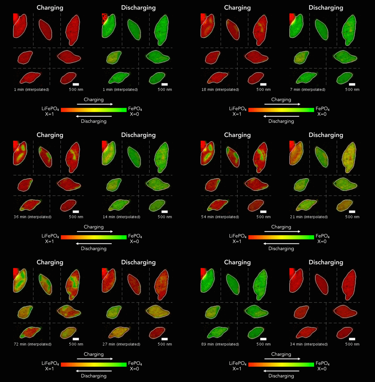
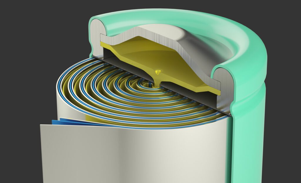
In pursuit of their quest for more capable batteries, scientists are searching for better ways to observe battery particles as they charge and discharge in real time. A new technique developed at Berkeley Lab’s Advanced Light Source is providing researchers with nanoscale images of the electrochemical reactions that make lithium-ion batteries do their thing.
In “Origin and hysteresis of lithium compositional spatiodynamics within battery primary particles,” published in Science, a team of researchers from the SLAC National Accelerator Laboratory, Berkeley Lab, Stanford University, et al describe their specially designed “liquid electrochemical STXM nanoimaging platform,” which uses soft X-rays to image lithium iron phosphate particles as they charge (delithiate) and discharge (lithiate) in a liquid electrolyte.
“The platform we developed allows us to image battery dynamics at the mesoscale, which is between a few nanometers and a few hundreds of nanometers,” says team leader Will Chueh. “This is a very difficult length scale to image in a functioning battery, but it’s critically important, because this is the scale that controls the fundamental processes involved in battery degradation and recharge time.”
“Our STXM-based platform provides the ability to image these electrochemical changes within a single battery particle,” adds team member David Shapiro. “And it offers the real-time resolution required to map the changes in the particles’ chemical composition and current densities, at a sub-particle scale, as the particles lithiate and delithiate.”
Previously, scientists have used transmission electron microscopy (TEM) to study working batteries at the nanoscale. This approach offers good spatial resolution, but X-ray microscopy can image a larger field of view and thicker materials than TEM, so it can study materials that more closely resemble real-world batteries.
Shapiro and colleagues are now building even more powerful X-ray microscopes, hoping to improve the platform’s spatial resolution by a factor of ten. “We are now working to achieve a spatial resolution approaching the soft X-ray wavelength, between one and five nanometers,” says Shapiro. “This will enable us to image chemical phases within the smallest available particles, often less than 100 nanometers in size, and still give us the penetrating power to look at large volumes of material, such as thousands of battery particles.”

